国际妇产科学杂志 ›› 2024, Vol. 51 ›› Issue (2): 220-227.doi: 10.12280/gjfckx.20231109
虞梁, 袁琳, 孟黄洋, 杨雨琴, 赵明睿, 张林, 程文俊( )
)
收稿日期:2023-12-29
出版日期:2024-04-15
发布日期:2024-04-19
通讯作者:
程文俊,E-mail:基金资助:
YU Liang, YUAN Lin, MENG Huang-yang, YANG Yu-qin, ZHAO Ming-rui, ZHANG Lin, CHENG Wen-jun()
Received:2023-12-29
Published:2024-04-15
Online:2024-04-19
Contact:
CHENG Wen-jun, E-mail: 摘要:
目的: 探讨影响腹壁子宫内膜异位症透明细胞癌变(abdominal wall endometriosis associated clear cell carcinoma,AWE-CCC)患者预后的相关因素。方法: 系统检索PubMed、EMBASE、MEDLINE、Google Scholar、Cochrane Library、万方、维普及中国知网数据库中包含AWE-CCC的相关文献,检索时间范围为1980年1月—2023年9月。使用Kaplan-Meier单因素生存分析及多因素Cox分析影响预后的因素,使用Apriori算法总结临床治疗规则。结果: 纳入68例AWE-CCC患者。年龄≤46岁、病灶直径>8 cm、病灶复发及复发间隔≤6个月是影响患者生存的不良预后因素(均P<0.05)。子宫切除对患者复发具有保护作用(HR=0.08,95%CI:0.01~0.81,P=0.03),且病灶切除联合子宫切除是强关联治疗规则。结论: 在临床实践中,应重视患者年龄和病灶大小,初始治疗应包含病灶切除及子宫切除为基础的联合手术治疗以减少术后复发。
虞梁, 袁琳, 孟黄洋, 杨雨琴, 赵明睿, 张林, 程文俊. 基于病例报道腹壁子宫内膜异位症透明细胞癌变相关预后因素分析[J]. 国际妇产科学杂志, 2024, 51(2): 220-227.
YU Liang, YUAN Lin, MENG Huang-yang, YANG Yu-qin, ZHAO Ming-rui, ZHANG Lin, CHENG Wen-jun. The Prognosis Factors Abdominal Wall Endometriosis Associated Clear Cell Carcinoma: A Pooled Analysis Based on Case Reports[J]. Journal of International Obstetrics and Gynecology, 2024, 51(2): 220-227.
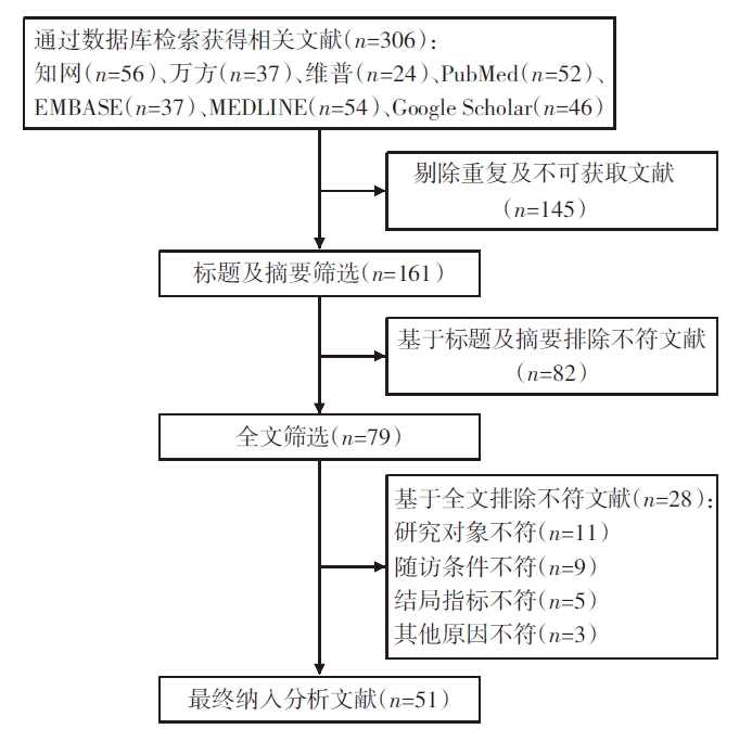
图1 文献筛选流程图
| 人群基本特征 | 报道情况 | n | M(P25,P75)或% |
|---|---|---|---|
| 患者年龄(岁) | 已知 | 68 | 46(42,53) |
| 病灶最大直径(cm) | 已知 | 57 | 9(6,13) |
| 未知 | 11 | — | |
| 病灶诊断延迟时间(年) | 已知 | 48 | 17(13,22) |
| 未知 | 20 | — | |
| 诊断恶变前CA125状态 | ≤35 U/mL | 30 | 44.2 |
| >35 U/mL | 19 | 27.9 | |
| 未知 | 19 | 27.9 | |
| AWE-CCC相关治疗方式 | |||
| 病灶切除 | 接受 | 66 | 97.1 |
| 未接受 | 2 | 2.9 | |
| 子宫切除 | 接受 | 44 | 64.7 |
| 未接受 | 24 | 35.3 | |
| 附件切除 | 接受 | 44 | 64.7 |
| 未接受 | 24 | 35.3 | |
| 大网膜切除 | 接受 | 23 | 33.8 |
| 未接受 | 45 | 66.2 | |
| 淋巴结切除 | 接受 | 22 | 32.4 |
| 未接受 | 46 | 67.6 | |
| 辅助化疗 | 接受 | 51 | 75.0 |
| 未接受 | 17 | 25.0 | |
| 辅助放疗 | 接受 | 19 | 27.9 |
| 未接受 | 49 | 72.1 | |
| 病灶切缘受累状态 | 阳性 | 6 | 8.8 |
| 阴性 | 24 | 35.3 | |
| 未知 | 38 | 55.9 | |
| 淋巴结受累状态 | 阳性 | 18 | 26.5 |
| 阴性 | 7 | 10.3 | |
| 未知 | 43 | 63.2 | |
| 恶变复发情况 | 复发 | 27 | 39.7 |
| 未复发 | 30 | 44.1 | |
| 未知 | 11 | 16.2 | |
| 生存情况 | 生存 | 54 | 79.4 |
| 死亡 | 14 | 20.6 |
表1 患者基本特征摘要表 (n=68)
| 人群基本特征 | 报道情况 | n | M(P25,P75)或% |
|---|---|---|---|
| 患者年龄(岁) | 已知 | 68 | 46(42,53) |
| 病灶最大直径(cm) | 已知 | 57 | 9(6,13) |
| 未知 | 11 | — | |
| 病灶诊断延迟时间(年) | 已知 | 48 | 17(13,22) |
| 未知 | 20 | — | |
| 诊断恶变前CA125状态 | ≤35 U/mL | 30 | 44.2 |
| >35 U/mL | 19 | 27.9 | |
| 未知 | 19 | 27.9 | |
| AWE-CCC相关治疗方式 | |||
| 病灶切除 | 接受 | 66 | 97.1 |
| 未接受 | 2 | 2.9 | |
| 子宫切除 | 接受 | 44 | 64.7 |
| 未接受 | 24 | 35.3 | |
| 附件切除 | 接受 | 44 | 64.7 |
| 未接受 | 24 | 35.3 | |
| 大网膜切除 | 接受 | 23 | 33.8 |
| 未接受 | 45 | 66.2 | |
| 淋巴结切除 | 接受 | 22 | 32.4 |
| 未接受 | 46 | 67.6 | |
| 辅助化疗 | 接受 | 51 | 75.0 |
| 未接受 | 17 | 25.0 | |
| 辅助放疗 | 接受 | 19 | 27.9 |
| 未接受 | 49 | 72.1 | |
| 病灶切缘受累状态 | 阳性 | 6 | 8.8 |
| 阴性 | 24 | 35.3 | |
| 未知 | 38 | 55.9 | |
| 淋巴结受累状态 | 阳性 | 18 | 26.5 |
| 阴性 | 7 | 10.3 | |
| 未知 | 43 | 63.2 | |
| 恶变复发情况 | 复发 | 27 | 39.7 |
| 未复发 | 30 | 44.1 | |
| 未知 | 11 | 16.2 | |
| 生存情况 | 生存 | 54 | 79.4 |
| 死亡 | 14 | 20.6 |
| 因素 | 2年累计死亡率[%(a/n)] | P | |
|---|---|---|---|
| 患者年龄 | ≤46岁 | 25.71(9/33) | 0.021 |
| >46岁 | 9.09(3/35) | ||
| 病灶最大直径 | ≤8 cm | 7.14(2/28) | 0.045 |
| >8 cm | 27.59(8/29) | ||
| 诊断恶变前CA125 | ≤35 U/mL | 16.67(5/30) | 0.138 |
| >35 U/mL | 26.32(5/19) | ||
| 病灶切缘受累状态 | 阳性 | 33.33(2/6) | 0.254 |
| 阴性 | 12.50(3/24) | ||
| 淋巴结受累状态 | 阳性 | 27.78(5/18) | 0.088 |
| 阴性 | 0.00(0/7) | ||
| 复发状态 | 复发 | 14.81(4/27) | 0.036 |
| 未复发 | 0.00(0/30) | ||
| DFS | ≤6个月 | 23.08(3/13) | 0.038 |
| >6个月 | 7.14(1/14) | ||
表2 单因素生存分析结果
| 因素 | 2年累计死亡率[%(a/n)] | P | |
|---|---|---|---|
| 患者年龄 | ≤46岁 | 25.71(9/33) | 0.021 |
| >46岁 | 9.09(3/35) | ||
| 病灶最大直径 | ≤8 cm | 7.14(2/28) | 0.045 |
| >8 cm | 27.59(8/29) | ||
| 诊断恶变前CA125 | ≤35 U/mL | 16.67(5/30) | 0.138 |
| >35 U/mL | 26.32(5/19) | ||
| 病灶切缘受累状态 | 阳性 | 33.33(2/6) | 0.254 |
| 阴性 | 12.50(3/24) | ||
| 淋巴结受累状态 | 阳性 | 27.78(5/18) | 0.088 |
| 阴性 | 0.00(0/7) | ||
| 复发状态 | 复发 | 14.81(4/27) | 0.036 |
| 未复发 | 0.00(0/30) | ||
| DFS | ≤6个月 | 23.08(3/13) | 0.038 |
| >6个月 | 7.14(1/14) | ||
| 因素 | P | HR(95%CI) | |
|---|---|---|---|
| 年龄 | ≤46岁 | - | - |
| >46岁 | 0.88 | 1.07(0.48~2.38) | |
| 子宫切除 | 否 | - | - |
| 是 | 0.03 | 0.08(0.01~0.81) | |
| 附件切除 | 否 | - | - |
| 是 | 0.06 | 9.64(0.92~100.97) | |
| 大网膜切除 | 否 | - | - |
| 是 | 0.99 | 1.00(0.38~2.66) | |
| 淋巴结切除 | 否 | - | - |
| 是 | 0.23 | 1.78(0.70~4.53) | |
| 辅助放疗 | 否 | - | - |
| 是 | 0.99 | 1.00(0.34~2.94) | |
| 辅助化疗 | 否 | - | - |
| 是 | 0.34 | 1.81(0.54~6.10) | |
表3 基于复发状态的多因素Cox分析结果
| 因素 | P | HR(95%CI) | |
|---|---|---|---|
| 年龄 | ≤46岁 | - | - |
| >46岁 | 0.88 | 1.07(0.48~2.38) | |
| 子宫切除 | 否 | - | - |
| 是 | 0.03 | 0.08(0.01~0.81) | |
| 附件切除 | 否 | - | - |
| 是 | 0.06 | 9.64(0.92~100.97) | |
| 大网膜切除 | 否 | - | - |
| 是 | 0.99 | 1.00(0.38~2.66) | |
| 淋巴结切除 | 否 | - | - |
| 是 | 0.23 | 1.78(0.70~4.53) | |
| 辅助放疗 | 否 | - | - |
| 是 | 0.99 | 1.00(0.34~2.94) | |
| 辅助化疗 | 否 | - | - |
| 是 | 0.34 | 1.81(0.54~6.10) | |
| 序号 | 前项数量 | 前项 | 后项 | 支持度(%) | 置信度(%) | 提升度 |
|---|---|---|---|---|---|---|
| 1 | 1 | 子宫切除 | 病灶切除 | 64.71 | 100.00 | 1.03 |
| 2 | 1 | 附件切除 | 病灶切除 | 64.71 | 100.00 | 1.03 |
| 3 | 1 | 子宫切除 | 附件切除 | 64.71 | 93.18 | 1.44 |
| 4 | 1 | 附件切除 | 子宫切除 | 64.71 | 93.18 | 1.44 |
| 5 | 1 | 附件切除 | 辅助化疗 | 64.71 | 81.82 | 1.09 |
| 6 | 2 | 子宫切除+病灶切除 | 附件切除 | 64.71 | 93.18 | 1.44 |
| 7 | 2 | 附件切除+病灶切除 | 子宫切除 | 64.71 | 93.18 | 1.44 |
| 8 | 2 | 附件切除+病灶切除 | 辅助化疗 | 64.71 | 81.82 | 1.09 |
| 9 | 2 | 子宫切除+附件切除 | 病灶切除 | 60.29 | 100.00 | 1.03 |
| 10 | 2 | 子宫切除+附件切除 | 辅助化疗 | 60.29 | 82.93 | 1.11 |
| 11 | 2 | 附件切除+辅助化疗 | 病灶切除 | 52.94 | 100.00 | 1.03 |
| 12 | 2 | 附件切除+辅助化疗 | 子宫切除 | 52.94 | 94.44 | 1.46 |
| 13 | 2 | 子宫切除+辅助化疗 | 病灶切除 | 51.47 | 100.00 | 1.03 |
| 14 | 2 | 子宫切除+辅助化疗 | 附件切除 | 51.47 | 97.14 | 1.50 |
| 15 | 3 | 子宫切除+附件切除+病灶切除 | 辅助化疗 | 60.29 | 82.93 | 1.11 |
| 16 | 3 | 附件切除+辅助化疗+病灶切除 | 子宫切除 | 52.94 | 94.44 | 1.46 |
| 17 | 3 | 子宫切除+辅助化疗+病灶切除 | 附件切除 | 51.47 | 97.14 | 1.50 |
| 18 | 3 | 子宫切除+附件切除+辅助化疗 | 病灶切除 | 50.00 | 100.00 | 1.03 |
表4 治疗方案关联规则分析结果
| 序号 | 前项数量 | 前项 | 后项 | 支持度(%) | 置信度(%) | 提升度 |
|---|---|---|---|---|---|---|
| 1 | 1 | 子宫切除 | 病灶切除 | 64.71 | 100.00 | 1.03 |
| 2 | 1 | 附件切除 | 病灶切除 | 64.71 | 100.00 | 1.03 |
| 3 | 1 | 子宫切除 | 附件切除 | 64.71 | 93.18 | 1.44 |
| 4 | 1 | 附件切除 | 子宫切除 | 64.71 | 93.18 | 1.44 |
| 5 | 1 | 附件切除 | 辅助化疗 | 64.71 | 81.82 | 1.09 |
| 6 | 2 | 子宫切除+病灶切除 | 附件切除 | 64.71 | 93.18 | 1.44 |
| 7 | 2 | 附件切除+病灶切除 | 子宫切除 | 64.71 | 93.18 | 1.44 |
| 8 | 2 | 附件切除+病灶切除 | 辅助化疗 | 64.71 | 81.82 | 1.09 |
| 9 | 2 | 子宫切除+附件切除 | 病灶切除 | 60.29 | 100.00 | 1.03 |
| 10 | 2 | 子宫切除+附件切除 | 辅助化疗 | 60.29 | 82.93 | 1.11 |
| 11 | 2 | 附件切除+辅助化疗 | 病灶切除 | 52.94 | 100.00 | 1.03 |
| 12 | 2 | 附件切除+辅助化疗 | 子宫切除 | 52.94 | 94.44 | 1.46 |
| 13 | 2 | 子宫切除+辅助化疗 | 病灶切除 | 51.47 | 100.00 | 1.03 |
| 14 | 2 | 子宫切除+辅助化疗 | 附件切除 | 51.47 | 97.14 | 1.50 |
| 15 | 3 | 子宫切除+附件切除+病灶切除 | 辅助化疗 | 60.29 | 82.93 | 1.11 |
| 16 | 3 | 附件切除+辅助化疗+病灶切除 | 子宫切除 | 52.94 | 94.44 | 1.46 |
| 17 | 3 | 子宫切除+辅助化疗+病灶切除 | 附件切除 | 51.47 | 97.14 | 1.50 |
| 18 | 3 | 子宫切除+附件切除+辅助化疗 | 病灶切除 | 50.00 | 100.00 | 1.03 |
| [1] |
Liu G, Wang Y, Chen Y, et al. Malignant transformation of abdominal wall endometriosis: A systematic review of the epidemiology, diagnosis, treatment, and outcomes[J]. Eur J Obstet Gynecol Reprod Biol, 2021, 264:363-367. doi: 10.1016/j.ejogrb.2021.08.006.
pmid: 34391052 |
| [2] | 陈颖, 乔媛, 王细文, 等. 腹壁子宫内膜异位症恶变为透明细胞癌1例报道[J]. 肿瘤防治研究, 2023, 50(5):544-546. doi: 10.3971/j.issn.1000-8578.2023.22.1061. |
| [3] | 周英凤, 顾莺, 胡雁, 等. JBI循证卫生保健中心关于不同类型研究的质量评价工具——病例报告及病例系列的质量评价[J]. 护士进修杂志, 2018, 33(4):310-312. doi: 10.16821/j.cnki.hsjx.2018.04.006. |
| [4] | Fassi E, Mandruzzato M, Zamparini M, et al. Clinical presentation and outcome of patients with enteric-type adenocarcinoma of the lung: A pooled analysis of published cases[J]. Lung Cancer, 2023, 179:107176. doi: 10.1016/j.lungcan.2023.107176. |
| [5] | Schnieber D, Wagner-Kolb D. Malignant transformation of extragenital endometriosis[J]. Geburtshilfe Frauenheilkd, 1986, 46(9):658-659. doi: 10.1055/s-2008-1036277. |
| [6] |
Hitti IF, Glasberg SS, Lubicz S. Clear cell carcinoma arising in extraovarian endometriosis: report of three cases and review of the literature[J]. Gynecol Oncol, 1990, 39(3):314-320. doi: 10.1016/0090-8258(90)90259-n.
pmid: 2258077 |
| [7] |
Ahn GH, Scully RE. Clear cell carcinoma of the inguinal region arising from endometriosis[J]. Cancer, 1991, 67(1):116-120. doi: 10.1002/1097-0142(19910101)67:1<116::aid-cncr2820670121>3.0.co;2-e
pmid: 1985706 |
| [8] |
Miller DM, Schouls JJ, Ehlen TG. Clear cell carcinoma arising in extragonadal endometriosis in a caesarean section scar during pregnancy[J]. Gynecol Oncol, 1998, 70(1):127-130. doi: 10.1006/gyno.1998.4989.
pmid: 9698489 |
| [9] |
Park SW, Hong SM, Wu HG, et al. Clear cell carcinoma arising in a Cesarean section scar endometriosis: a case report[J]. J Korean Med Sci, 1999, 14(2):217-219. doi: 10.3346/jkms.1999.14.2.217.
pmid: 10331572 |
| [10] | Klein AE, Bauer TW, Marks KE, et al. Papillary clear cell adenocarcinoma of the groin arising from endometriosis[J]. Clin Orthop Relat Res, 1999(361):192-198. doi: 10.1097/00003086-199904000-00025. |
| [11] |
Ishida GM, Motoyama T, Watanabe T, et al. Clear cell carcinoma arising in a cesarean section scar. Report of a case with fine needle aspiration cytology[J]. Acta Cytol, 2003, 47(6):1095-1098. doi: 10.1159/000326655.
pmid: 14674088 |
| [12] | Li JY, Chen YJ, Wu YC, et al. Two- and three-dimensional Doppler ultrasound analysis of abdominal wall clear cell carcinoma[J]. Ultrasound Obstet Gynecol, 2003, 22(1):98-100. doi: 10.1002/uog.173. |
| [13] | Sergent F, Baron M, Le Cornec JB, et al. Malignant transformation of abdominal wall endometriosis: a new case report[J]. J Gynecol Obstet Biol Reprod(Paris), 2006, 35(2):186-190. doi: 10.1016/s0368-2315(06)76394-3. |
| [14] | Alberto VO, Lynch M, Labbei FN, et al. Primary abdominal wall clear cell carcinoma arising in a Caesarean section scar endometriosis[J]. Ir J Med Sci, 2006, 175(1):69-71. doi: 10.1007/BF03169006. |
| [15] |
Razzouk K, Roman H, Chanavaz-Lacheray I, et al. Mixed clear cell and endometrioid carcinoma arising in parietal endometriosis[J]. Gynecol Obstet Invest, 2007, 63(3):140-142. doi: 10.1159/000096437.
pmid: 17057400 |
| [16] |
Harry VN, Shanbhag S, Lyall M, et al. Isolated clear cell adenocarcinoma in scar endometriosis mimicking an incisional hernia[J]. Obstet Gynecol, 2007, 110(2 Pt 2):469-471. doi: 10.1097/01.AOG.0000260393.46154.5f.
pmid: 17666631 |
| [17] | Bats AS, Zafrani Y, Pautier P, et al. Malignant transformation of abdominal wall endometriosis to clear cell carcinoma: case report and review of the literature[J]. Fertil Steril, 2008, 90(4):1197.e13-e16. doi: 10.1016/j.fertnstert.2007.08.080. |
| [18] | Achach T, Rammeh S, Trabelsi A, et al. Clear cell adenocarcinoma arising from abdominal wall endometriosis[J]. J Oncol, 2008, 2008:478325. doi: 10.1155/2008/478325. |
| [19] |
Matsuo K, Alonsozana EL, Eno ML, et al. Primary peritoneal clear cell adenocarcinoma arising in previous abdominal scar for endometriosis surgery[J]. Arch Gynecol Obstet, 2009, 280(4):637-641. doi: 10.1007/s00404-009-0962-y.
pmid: 19219617 |
| [20] |
Williams C, Petignat P, Belisle A, et al. Primary abdominal wall clear cell carcinoma: case report and review of literature[J]. Anticancer Res, 2009, 29(5):1591-1593.
pmid: 19443371 |
| [21] | Bourdel N, Durand M, Gimbergues P, et al. Exclusive nodal recurrence after treatment of degenerated parietal endometriosis[J]. Fertil Steril, 2010, 93(6):2074.e1-e6. doi: 10.1016/j.fertnstert.2009.11.004. |
| [22] | 颜耀华, 李力, 郭建新, 等. 剖宫产术后腹壁瘢痕子宫内膜异位症癌变一例[J]. 中华妇产科杂志, 2010, 45(4):319. doi: 10.3760/cma.j.issn.0529-567x.2010.04.022. |
| [23] |
Yan Y, Li L, Guo J, et al. Malignant transformation of an endometriotic lesion derived from an abdominal wall scar[J]. Int J Gynaecol Obstet, 2011, 115(2):202-203. doi: 10.1016/j.ijgo.2011.06.018.
pmid: 21872248 |
| [24] |
Li X, Yang J, Cao D, et al. Clear-cell carcinoma of the abdominal wall after cesarean delivery[J]. Obstet Gynecol, 2012, 120(<W>2 Pt 2):445-448. doi: 10.1097/AOG.0b013e31824da6fe.
pmid: 22825261 |
| [25] | Mert I, Semaan A, Kim S, et al. Clear cell carcinoma arising in the abdominal wall: two case reports and literature review[J]. Am J Obstet Gynecol, 2012, 207(2):e7-e9. doi: 10.1016/j.ajog.2012.05.029. |
| [26] |
Shalin SC, Haws AL, Carter DG, et al. Clear cell adenocarcinoma arising from endometriosis in abdominal wall cesarean section scar: a case report and review of the literature[J]. J Cutan Pathol, 2012, 39(11):1035-1041. doi: 10.1111/j.1600-0560.2012.01982.x.
pmid: 22882475 |
| [27] | Sawazaki H, Goto H, Takao N, et al. Clear cell adenocarcinoma arising from abdominal wall endometriosis mimicking urachal tumor[J]. Urology, 2012, 79(6):e84-e85. doi: 10.1016/j.urology.2011.02.034. |
| [28] | 江茂情, 孙龙, 吴华. 18F-FDG PET/CT诊断腹壁子宫内膜异位恶变术后复发1例并文献复习[J]. 中国临床医学影像杂志, 2013, 24(4):301-303. doi: 10.3969/j.issn.1008-1062.2013.04.026. |
| [29] | Heller DS, Houck K, Lee ES, et al. Clear cell adenocarcinoma of the abdominal wall: a case report[J]. J Reprod Med, 2014, 59(5/6):330-332. |
| [30] | Ijichi S, Mori T, Suganuma I, et al. Clear cell carcinoma arising from cesarean section scar endometriosis: case report and review of the literature[J]. Case Rep Obstet Gynecol, 2014, 2014:642483. doi: 10.1155/2014/642483. |
| [31] |
Liu H, Leng J, Lang J, et al. Clear cell carcinoma arising from abdominal wall endometriosis: a unique case with bladder and lymph node metastasis[J]. World J Surg Oncol, 2014, 12:51. doi: 10.1186/1477-7819-12-51.
pmid: 24597651 |
| [32] | Aust S, Tiringer D, Grimm C, et al. Therapy of a clear cell adenocarcinoma of unknown primary arising in the abdominal wall after cesarean section and after hysterectomy[J]. Wien Klin Wochenschr, 2015, 127(1/2):62-64. doi: 10.1007/s00508-014-0619-0. |
| [33] | Ruiz MP, Wallace DL, Connell MT. Transformation of Abdominal Wall Endometriosis to Clear Cell Carcinoma[J]. Case Rep Obstet Gynecol, 2015, 2015:123740. doi: 10.1155/2015/123740. |
| [34] |
Sosa-Durán EE, Aboharp-Hasan Z, Mendoza-Morales RC, et al. Clear cell adenocarcinoma arising from abdominal wall endometriosis[J]. Cir Cir, 2016, 84(3):245-249. doi: 10.1016/j.circir.2015.06.024.
pmid: 26272425 |
| [35] |
Ferrandina G, Palluzzi E, Fanfani F, et al. Endometriosis-associated clear cell carcinoma arising in caesarean section scar: a case report and review of the literature[J]. World J Surg Oncol, 2016, 14(1):300. doi: 10.1186/s12957-016-1054-7.
pmid: 27912770 |
| [36] |
Kostrzeba E, Barczyk M, Wichtowski M, et al. Clear Cell Carcinoma of the abdominal wall[J]. Pol Przegl Chir, 2017, 89(6):40-43. doi: 10.5604/01.3001.0010.6753.
pmid: 29335387 |
| [37] | Marques C, Silva TS, Dias MF. Clear cell carcinoma arising from abdominal wall endometriosis-Brief report and review of the literature[J]. Gynecol Oncol Rep, 2017, 20:78-80. doi: 10.1016/j.gore.2017.03.008. |
| [38] |
Gentile JKA, Migliore R, Kistenmacker FJN, et al. Malignant transformation of abdominal wall endometriosis to clear cell carcinoma: case report[J]. Sao Paulo Med J, 2018, 136(6):586-590. doi: 10.1590/1516-3180.2017.0103300417.
pmid: 29116312 |
| [39] | Yoshida S, Onogi A, Kuwahara M, et al. Clear Cell Adenocarcinoma Arising from Endometriosis in the Groin: Wide Resection and Reconstruction with a Fascia Lata Tensor Muscle Skin Flap[J]. Case Rep Obstet Gynecol, 2018, 2018:2139595. doi: 10.1155/2018/2139595. |
| [40] | 张承, 赵昊云, 钱敏, 等. 腹壁切口子宫内膜异位症恶变为透明细胞癌一例[J]. 中华妇产科杂志, 2018, 53(12):869. doi: 10.3760/cma.j.issn.0529-567x.2018.12.015. |
| [41] | Lai YL, Hsu HC, Kuo KT, et al. Clear Cell Carcinoma of the Abdominal Wall as a Rare Complication of General Obstetric and Gynecologic Surgeries: 15 Years of Experience at a Large Academic Institution[J]. Int J Environ Res Public Health, 2019, 16(4):552. doi: 10.3390/ijerph16040552. |
| [42] | Tsuruga T, Hirata T, Akiyama I, et al. Mixed endometrioid and clear cell carcinoma arising from laparoscopic trocar site endometriosis[J]. J Obstet Gynaecol Res, 2019, 45(8):1613-1618. doi: 10.1111/jog.14014. |
| [43] |
Lopes A, Anton C, Slomovitz BM, et al. Clear cell carcinoma arising from abdominal wall endometrioma after cesarean section[J]. Int J Gynecol Cancer, 2019, 29(8):1332-1335. doi: 10.1136/ijgc-2019-000808.
pmid: 31451559 |
| [44] | Provendier A, Angeles MA, Meyrignac O, et al. Clear cell adenocarcinoma arising from the abdominal wall after cesarean section in a patient with uterine adenomyosis[J]. J Surg Case Rep, 2020, 2020(4): rjaa070. doi: 10.1093/jscr/rjaa070. |
| [45] |
Giannella L, Serri M, Maccaroni E, et al. Endometriosis-associated Clear Cell Carcinoma of the Abdominal Wall After Caesarean Section: A Case Report and Review of the Literature[J]. In Vivo, 2020, 34(4):2147-2152. doi: 10.21873/invivo.12021.
pmid: 32606196 |
| [46] | Colarossi C, Picardo MC, Colarossi L, et al. Clear Cell Carcinoma Arising in an Abdominal Wall Cesarean Section Scar: A Case Report With Description of Pathological and Molecular Features[J]. Front Surg, 2021, 8:735381. doi: 10.3389/fsurg.2021.735381. |
| [47] | Kadam SS, Deshmukh S, Karandikar SM, et al. Does Preoperative Diagnosis Will Change the Treatment Plan of Clear Cell Carcinoma of Endometrium Masquerading as Desmoid Tumor of Anterior Abdominal Wall?: a Case Report[J]. Indian J Surg Oncol, 2021, 12(Suppl 2):312-318. doi: 10.1007/s13193-021-01352-2. |
| [48] | 杨泽成, 史常赛, 陈佳乐, 等. 腹壁子宫内膜异位症相关性透明细胞癌一例及文献回顾[J]. 中华疝和腹壁外科杂志(电子版), 2022, 16(5):595-599. doi: 10.3877/cma.j.issn.1674-392X.2022.05.023. |
| [49] | 孙国栋, 姜旖, 袁琳, 等. 巨大腹壁子宫内膜异位症恶变一例并文献复习[J]. 国际妇产科学杂志, 2022, 49(3):321-324. doi: 10.12280/gjfckx.20210944. |
| [50] |
Feng Z, Wen H, Ju X, et al. Treatment for clear cell carcinoma of the abdominal wall at a tertiary cancer center[J]. Sci Rep, 2022, 12(1):10820. doi: 10.1038/s41598-022-14917-0.
pmid: 35752641 |
| [51] |
Bahall V, De Barry L, Rampersad A. Clear cell carcinoma arising from abdominal wall endometriosis-a report on two cases and literature review[J]. World J Surg Oncol, 2022, 20(1):86. doi: 10.1186/s12957-022-02553-x.
pmid: 35292079 |
| [52] | Lewis GK, Gajarawala SN, Robinson KE, et al. Clear cell adenocarcinoma arising from anterior abdominal wall cesarean section scar endometriosis treated with excision and the addition of Trastuzumab for adjuvant chemotherapy: A case report[J]. Gynecol Oncol Rep, 2022, 41:100995. doi: 10.1016/j.gore.2022.100995. |
| [53] | Bellalah A, Lahbecha B, Zokar O, et al. Clear cell carcinoma of the abdominal wall: A case report with a review of the literature[J]. Ann Med Surg(Lond), 2022, 79:104038. doi: 10.1016/j.amsu.2022.104038. |
| [54] |
Liu D, Wei H, Huang J, et al. Clear Cell Adenocarcinoma Arising from Endometriosis in Abdominal Wall Cesarean Section Scar: A Case Report and Literature Review[J]. Int J Womens Health, 2023, 15:25-32. doi: 10.2147/IJWH.S382235.
pmid: 36636515 |
| [55] | Li Y, Xiu L, Ma M, et al. Developing and validating a prognostic nomogram for ovarian clear cell carcinoma patients: A retrospective comparison of lymph node staging schemes with competing risk analysis[J]. Front Oncol, 2022, 12:940601. doi: 10.3389/fonc.2022.940601. |
| [56] |
Zhu C, Zhu J, Qian L, et al. Clinical characteristics and prognosis of ovarian clear cell carcinoma: a 10-year retrospective study[J]. BMC Cancer, 2021, 21(1):322. doi: 10.1186/s12885-021-08061-7.
pmid: 33766002 |
| [57] | Cao BY, Tong F, Zhang LT, et al. Risk factors, prognostic predictors, and nomograms for pancreatic cancer patients with initially diagnosed synchronous liver metastasis[J]. World J Gastrointest Oncol, 2023, 15(1):128-142. doi: 10.4251/wjgo.v15.i1.128. |
| [58] |
Li Z, Duan H, Guo J, et al. Discussion on the rationality of FIGO 2018 stage IIIC for cervical cancer with oncological outcomes: a cohort study[J]. Ann Transl Med, 2022, 10(2):122. doi: 10.21037/atm-21-6374.
pmid: 35282078 |
| [59] |
Deng S, Jiang Z, Cao Y, et al. Development and validation of a prognostic scoring system for patients with colorectal cancer hepato-pulmonary metastasis: a retrospective study[J]. BMC Cancer, 2022, 22(1):643. doi: 10.1186/s12885-022-09738-3.
pmid: 35690752 |
| [60] | Liao Z, Wang D, Song N, et al. Prognosis of clear cell renal cell carcinoma patients stratified by age: A research relied on SEER database[J]. Front Oncol, 2022, 12:975779. doi: 10.3389/fonc.2022.975779. |
| [61] |
Yang H, Ji X, Jin C, et al. A Practical Nomogram for Predicting the Prognosis of Elderly Patients with Gastric Adenocarcinoma After Gastrectomy[J]. Int J Gen Med, 2022, 15:473-488. doi: 10.2147/IJGM.S343306.
pmid: 35046708 |
| [62] |
Taburiaux L, Pluchino N, Petignat P, et al. Endometriosis-Associated Abdominal Wall Cancer: A Poor Prognosis?[J]. Int J Gynecol Cancer, 2015, 25(9):1633-1638. doi: 10.1097/IGC.0000000000000556.
pmid: 26448542 |
| [63] |
Zhou Y, Hua KQ. Ovarian endometriosis: risk factor analysis and prediction of malignant transformation[J]. Prz Menopauzalny, 2018, 17(1):43-48. doi: 10.5114/pm.2018.74902.
pmid: 29725285 |
| [64] |
Shinmura H, Yoneyama K, Harigane E, et al. Use of tumor markers to distinguish endometriosis-related ovarian neoplasms from ovarian endometrioma[J]. Int J Gynecol Cancer, 2020, 30(6):831-836. doi: 10.1136/ijgc-2020-001210.
pmid: 32354795 |
| [65] | Li M, Tan J, Zhang Y, et al. Assessing CT imaging features combined with CEA and CA125 levels to identify endometriosis-associated ovarian cancer[J]. Abdom Radiol(NY), 2021, 46(6):2367-2375. doi: 10.1007/s00261-020-02571-x. |
| [66] |
Bai H, Sha G, Xiao M, et al. The prognostic value of pretreatment CA-125 levels and CA-125 normalization in ovarian clear cell carcinoma: a two-academic-institute study[J]. Oncotarget, 2016, 7(13):15566-15576. doi: 10.18632/oncotarget.7216.
pmid: 26863639 |
| [67] |
Gundogdu F, Soylu F, Erkan L, et al. The role of serum CA-125 levels and CA-125 tissue expression positivity in the prediction of the recurrence of stage Ⅲ and Ⅳ epithelial ovarian tumors (CA-125 levels and tissue CA-125 in ovarian tumors)[J]. Arch Gynecol Obstet, 2011, 283(6):1397-1402. doi: 10.1007/s00404-010-1589-8.
pmid: 20645105 |
| [68] |
Colombo N, Sessa C, du Bois A, et al. ESMO-ESGO consensus conference recommendations on ovarian cancer: pathology and molecular biology, early and advanced stages, borderline tumours and recurrent disease[J]. Ann Oncol, 2019, 30(5):672-705. doi: 10.1093/annonc/mdz062.
pmid: 31987337 |
| [69] | 钱欢欢, 戴辉华, 林明娟, 等. 子宫内膜异位症相关恶性肿瘤的研究进展[J]. 南京医科大学学报(自然科学版), 2019, 39(3):442-447. doi: 10.7655/NYDXBNS20190327. |
| [70] |
Crotzer DR, Sun CC, Coleman RL, et al. Lack of effective systemic therapy for recurrent clear cell carcinoma of the ovary[J]. Gynecol Oncol, 2007, 105(2):404-408. doi: 10.1016/j.ygyno.2006.12.024.
pmid: 17292461 |
| [1] | 曹秀蓉, 周文柏, 范香, 王逸斐, 朱鹏峰. 单细胞RNA测序解析子宫内膜异位症血管生成机制[J]. 国际妇产科学杂志, 2025, 52(2): 199-205. |
| [2] | 殷婷, 丛慧芳. 子宫内膜异位症与痛觉敏化的免疫学研究进展[J]. 国际妇产科学杂志, 2025, 52(2): 206-210. |
| [3] | 江爱美, 张信美. 腹壁子宫内膜异位症的治疗进展[J]. 国际妇产科学杂志, 2025, 52(2): 211-216. |
| [4] | 白耀俊, 王思瑶, 令菲菲, 张森淮, 李红丽, 刘畅. Trop-2及靶向Trop-2抗体偶联药物在妇科恶性肿瘤中的应用进展[J]. 国际妇产科学杂志, 2025, 52(1): 1-7. |
| [5] | 张云凤, 张宛玥, 卢悦, 王阳阳, 井佳雨, 牟婧祎, 王悦. ARID1A与PIK3CA突变在卵巢子宫内膜异位症恶变中的研究进展[J]. 国际妇产科学杂志, 2025, 52(1): 19-22. |
| [6] | 石百超, 王宇, 常惠, 卢凤娟, 关木馨, 余健楠, 吴效科. 中药及天然产物改善子宫内膜异位症的作用机制[J]. 国际妇产科学杂志, 2025, 52(1): 66-71. |
| [7] | 豆苗苗, 郑婧, 张航, 杨博, 张春洁, 刘志杰. 子宫附腔畸形的诊断及预后分析一例[J]. 国际妇产科学杂志, 2025, 52(1): 84-88. |
| [8] | 郭希, 刘思敏, 魏佳, 杨永秀. 卵巢及输卵管子宫内膜异位症恶变为透明细胞癌一例[J]. 国际妇产科学杂志, 2024, 51(6): 680-683. |
| [9] | 张艳, 张意茗. 子宫腺肌病合并卵巢子宫内膜异位囊肿取卵术后并发盆腔脓肿一例[J]. 国际妇产科学杂志, 2024, 51(6): 717-720. |
| [10] | 李慧敏, 胡雅莉, 张森淮, 马晓梅, 许飞雪. 高强度聚焦超声技术在妇产科疾病中的应用进展[J]. 国际妇产科学杂志, 2024, 51(5): 486-491. |
| [11] | 何清, 胡红波. 人工智能在子宫内膜癌诊治中的应用与展望[J]. 国际妇产科学杂志, 2024, 51(5): 572-577. |
| [12] | 季安璇, 赵淑华. 卵巢成熟性囊性畸胎瘤恶性转化一例[J]. 国际妇产科学杂志, 2024, 51(4): 384-387. |
| [13] | 郭希, 魏佳, 杨永秀. 导致子宫内膜疾病的激素通路及调节因素[J]. 国际妇产科学杂志, 2024, 51(4): 395-400. |
| [14] | 黄玥, 谭欣林, 袁玉红, 张强, 任郁, 宾冬梅, 漆洪波, 石琪. 四川经济欠发达地区围产期抑郁症状检出率及影响因素[J]. 国际妇产科学杂志, 2024, 51(4): 417-423. |
| [15] | 吴晓莉, 刘开江. 子宫内膜癌TCGA分子分型与治疗新进展[J]. 国际妇产科学杂志, 2024, 51(3): 247-252. |
| 阅读次数 | ||||||
|
全文 |
|
|||||
|
摘要 |
|
|||||