国际妇产科学杂志 ›› 2022, Vol. 49 ›› Issue (4): 407-410.doi: 10.12280/gjfckx.20220022

• 妇科肿瘤研究:病例报告 • 上一篇    下一篇

腹腔弥漫型平滑肌瘤病一例

张昊萌, 张洪扬, 邵红颖, 王艳秋(), 李长忠   

  1. 250021 济南,山东大学附属省立医院妇科(张昊萌,张洪扬,李长忠),麻醉手术科(王艳秋);山东中医药大学(邵红颖)
  • 收稿日期:2022-01-11 出版日期:2022-08-15 发布日期:2022-08-19
  • 通讯作者: 王艳秋 E-mail:540695769@qq.com
  • 基金资助:
    国家自然科学基金(81671434)

Leiomyomatosis Peritonealis Disseminate: A Case Report

ZHANG Hao-meng, ZHANG Hong-yang, SHAO Hong-ying, WANG Yan-qiu(), LI Chang-zhong   

  1. Department of Gynecology (ZHANG Hao-meng, ZHANG Hong-yang, LI Chang-zhong), Department of Anesthesia Surgery (WANG Yan-qiu), Shandong Provincial Hospital Affiliated to Shandong University, Jinan 250021, China;Shandong University of Traditional Chinese Medicine, Jinan 250355, China (SHAO Hong-ying)
  • Received:2022-01-11 Published:2022-08-15 Online:2022-08-19
  • Contact: WANG Yan-qiu E-mail:540695769@qq.com

摘要:

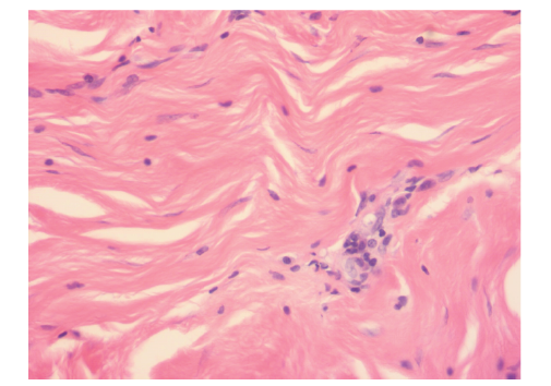
腹腔弥漫型平滑肌瘤病是一种罕见的特殊类型子宫平滑肌瘤,目前,其发病机制尚不明确,发病率极低,误诊率较高。报道山东大学附属省立医院2021年收治的1例腹腔弥漫型平滑肌瘤病患者,该患者以月经频发伴经量增多为首发症状,术前未明确诊断为腹腔弥漫型平滑肌瘤病,相关辅助检查也无明确指征,术中探查可见多枚平滑肌瘤样结节弥漫生长于腹腔。腹腔弥漫型平滑肌瘤病的病灶可弥漫分布于盆腹腔多器官表面,肉眼较难与腹膜转移癌或胃肠道间质肿瘤相鉴别。另外,在临床表现上,症状多与病灶种植位置有关,无特异性表现。在实际临床实践中,术前检出率极低,为降低误诊率,应对有多次子宫平滑肌瘤复发病史、腹腔镜碎瘤史的患者提高警惕,必要时行盆腹腔磁共振成像完善术前检查。

关键词: 平滑肌瘤病, 平滑肌瘤, 妇科外科手术, 磁共振成像, 病例报告

Abstract:

Leiomyomatosis peritonealis disseminate (LPD) is a rare special type of uterine leiomyoma. At present, the pathogenesis is not clear, its incidence is extremely low, and the misdiagnosis rate is high. A case of abdominal diffuse leiomyoma admitted to Shandong Provincial Hospital Affiliated to Shandong University in 2021 was reported. The first symptom of this patient was frequent menstruation with increased menstrual flow. Before operation, it was not clearly diagnosed as LPD, and there was no clear indication from related auxiliary examinations. During the operation, it was found that many leiomyoma-like nodules of uterine fibroids diffuse in the abdominal cavity. LPD can be diffusely distributed on the surfaces of pelvic and abdominal organs, and it is difficult to distinguish it from peritoneal metastatic carcinoma or gastrointestinal stromal tumor with naked eyes. In addition, the symptoms are mostly related to the location of the lesion, and there is no specific manifestation. In practical clinical practice, the preoperative detection rate is extremely low. In order to reduce the misdiagnosis rate, patients with multiple history of uterine leiomyoma and laparoscopically crushed tumor should be alert, and if necessary, preoperative examination of pelvic and abdominal magnetic resonance imaging should be performed.

Key words: Leiomyomatosis, Leiomyoma, Gynecologic surgical procedures, Magnetic resonance imaging, Case reports