| [1] |
Hooker AB, de Leeuw RA, Twisk J, et al. Reproductive performance of women with and without intrauterine adhesions following recurrent dilatation and curettage for miscarriage: long-term follow-up of a randomized controlled trial[J]. Hum Reprod, 2021, 36(1):70-81. doi: 10.1093/humrep/deaa289.
|
| [2] |
Lee WL, Liu CH, Cheng M, et al. Focus on the Primary Prevention of Intrauterine Adhesions: Current Concept and Vision[J]. Int J Mol Sci, 2021, 22(10):5175. doi: 10.3390/ijms22105175.
|
| [3] |
Qiao J, Wang Y, Li X, et al. A Lancet Commission on 70 years of women′s reproductive, maternal, newborn, child, and adolescent health in China[J]. Lancet, 2021, 397(10293):2497-2536. doi: 10.1016/S0140-6736(20)32708-2.
pmid: 34043953
|
| [4] |
Trinh TT, Nguyen KD, Pham HV, et al. Effectiveness of Hyaluronic Acid Gel and Intrauterine Devices in Prevention of Intrauterine Adhesions after Hysteroscopic Adhesiolysis in Infertile Women[J]. J Minim Invasive Gynecol, 2022, 29(2):284-290. doi: 10.1016/j.jmig.2021.08.010.
|
| [5] |
Song YT, Liu PC, Tan J, et al. Stem cell-based therapy for ameliorating intrauterine adhesion and endometrium injury[J]. Stem Cell Res Ther, 2021, 12(1):556. doi: 10.1186/s13287-021-02620-2.
|
| [6] |
Abudukeyoumu A, Li MQ, Xie F. Transforming growth factor-β1 in intrauterine adhesion[J]. Am J Reprod Immunol, 2020, 84(2):e13262. doi: 10.1111/aji.13262.
|
| [7] |
Rajabi N, Rezaei A, Kharaziha M, et al. Recent Advances on Bioprinted Gelatin Methacrylate-Based Hydrogels for Tissue Repair[J]. Tissue Eng Part A, 2021, 27(11/12):679-702. doi: 10.1089/ten.TEA.2020.0350.
|
| [8] |
Janarthanan G, Kim JH, Kim I, et al. Manufacturing of self-standing multi-layered 3D-bioprinted alginate-hyaluronate constructs by controlling the cross-linking mechanisms for tissue engineering applications[J]. Biofabrication, 2022 May 31;14(3). doi: 10.1088/1758-5090/ac6c4c.
|
| [9] |
Cai Y, Wu F, Yu Y, et al. Porous scaffolds from droplet microfluidics for prevention of intrauterine adhesion[J]. Acta Biomater, 2019, 84:222-230. doi: 10.1016/j.actbio.2018.11.016.
pmid: 30476581
|
| [10] |
Ji W, Hou B, Lin W, et al. 3D Bioprinting a human iPSC-derived MSC-loaded scaffold for repair of the uterine endometrium[J]. Acta Biomater, 2020, 116:268-284. doi: 10.1016/j.actbio.2020.09.012.
pmid: 32911103
|
| [11] |
Wang L, Yu C, Chang T, et al. In situ repair abilities of human umbilical cord-derived mesenchymal stem cells and autocrosslinked hyaluronic acid gel complex in rhesus monkeys with intrauterine adhesion[J]. Sci Adv, 2020, 6(21):eaba6357. doi: 10.1126/sciadv.aba6357.
|
| [12] |
Xin L, Lin X, Pan Y, et al. A collagen scaffold loaded with human umbilical cord-derived mesenchymal stem cells facilitates endometrial regeneration and restores fertility[J]. Acta Biomater, 2019, 92:160-171. doi: 10.1016/j.actbio.2019.05.012.
pmid: 31075515
|
| [13] |
Wenbo Q, Lijian X, Shuangdan Z, et al. Controlled releasing of SDF-1α in chitosan-heparin hydrogel for endometrium injury healing in rat model[J]. Int J Biol Macromol, 2020, 143:163-172. doi: 10.1016/j.ijbiomac.2019.11.184.
pmid: 31765745
|
| [14] |
Xu T, Jin J, Gregory C, et al. Inkjet printing of viable mammalian cells[J]. Biomaterials, 2005, 26(1):93-99. doi: 10.1016/j.biomaterials.2004.04.011.
pmid: 15193884
|
| [15] |
Matai I, Kaur G, Seyedsalehi A, et al. Progress in 3D bioprinting technology for tissue/organ regenerative engineering[J]. Biomaterials, 2020,226:119536. doi: 10.1016/j.biomaterials.2019.119536.
|
| [16] |
Melchels FP, Feijen J, Grijpma DW. A review on stereolithography and its applications in biomedical engineering[J]. Biomaterials, 2010, 31(24):6121-6130. doi: 10.1016/j.biomaterials.2010.04.050.
pmid: 20478613
|
| [17] |
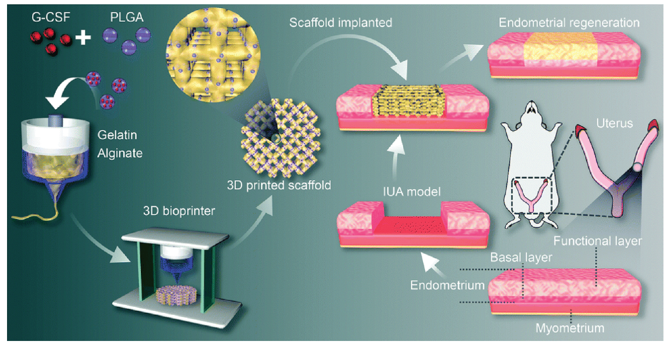
Wen J, Hou B, Lin W, et al. 3D-printed hydrogel scaffold-loaded granulocyte colony-stimulating factor sustained-release microspheres and their effect on endometrial regeneration[J]. Biomater Sci, 2022, 10(12):3346-3358. doi: 10.1039/d2bm00109h.
|
| [18] |
Min J, Lu N, Huang S, et al. Phenotype and biological characteristics of endometrial mesenchymal stem/stromal cells: A comparison between intrauterine adhesion patients and healthy women[J]. Am J Reprod Immunol, 2021, 85(6):e13379. doi: 10.1111/aji.13379.
|
| [19] |
Xiang E, Han B, Zhang Q, et al. Human umbilical cord-derived mesenchymal stem cells prevent the progression of early diabetic nephropathy through inhibiting inflammation and fibrosis[J]. Stem Cell Res Ther, 2020, 11(1):336. doi: 10.1186/s13287-020-01852-y.
pmid: 32746936
|
| [20] |
Gargett CE, Ye L. Endometrial reconstruction from stem cells[J]. Fertil Steril, 2012, 98(1):11-20. doi: 10.1016/j.fertnstert.2012.05.004.
pmid: 22657248
|
| [21] |
Zheng JH, Zhang JK, Kong DS, et al. Quantification of the CM-Dil-labeled human umbilical cord mesenchymal stem cells migrated to the dual injured uterus in SD rat[J]. Stem Cell Res Ther, 2020, 11(1):280. doi: 10.1186/s13287-020-01806-4.
|
| [22] |
Xu L, Ding L, Wang L, et al. Umbilical cord-derived mesenchymal stem cells on scaffolds facilitate collagen degradation via upregulation of MMP-9 in rat uterine scars[J]. Stem Cell Res Ther, 2017, 8(1):84. doi: 10.1186/s13287-017-0535-0.
pmid: 28420433
|
| [23] |
Cao Y, Sun H, Zhu H, et al. Allogeneic cell therapy using umbilical cord MSCs on collagen scaffolds for patients with recurrent uterine adhesion: a phaseⅠclinical trial[J]. Stem Cell Res Ther, 2018, 9(1):192. doi: 10.1186/s13287-018-0904-3.
|
| [24] |
Wei X, Liu F, Zhang S, et al. Human Umbilical Cord Mesenchymal Stem Cell-Derived Conditioned Medium Promotes Human Endometrial Cell Proliferation through Wnt/β-Catenin Signaling[J]. Biomed Res Int, 2022,2022:8796093. doi: 10.1155/2022/8796093.
|
| [25] |
Zhao YX, Chen SR, Huang QY, et al. Repair abilities of mouse autologous adipose-derived stem cells and ShakeGelTM3D complex local injection with intrauterine adhesion by BMP7-Smad5 signaling pathway activation[J]. Stem Cell Res Ther, 2021, 12(1):191. doi: 10.1186/s13287-021-02258-0.
|
| [26] |
Fonseca AC, Melchels F, Ferreira M, et al. Emulating Human Tissues and Organs: A Bioprinting Perspective Toward Personalized Medicine[J]. Chem Rev, 2020, 120(19):11128-11174. doi: 10.1021/acs.chemrev.0c00342.
|
| [27] |
Feng M, Hu S, Qin W, et al. Bioprinting of a Blue Light-Cross-Linked Biodegradable Hydrogel Encapsulating Amniotic Mesenchymal Stem Cells for Intrauterine Adhesion Prevention[J]. ACS Omega, 2021, 6(36):23067-23075. doi: 10.1021/acsomega.1c02117.
pmid: 34549107
|
| [28] |
Lu S, Wang XC, Li WZ, et al. Injectable 3D-Printed Porous Scaffolds for Adipose Stem Cell Delivery and Endometrial Regeneration[J]. Adv Funct Mater, 2023, 33(34):2303368. doi: 10.1002/adfm.202303368.
|
| [29] |
Ma T, Fu B, Yang X, et al. Adipose mesenchymal stem cell-derived exosomes promote cell proliferation, migration, and inhibit cell apoptosis via Wnt/β-catenin signaling in cutaneous wound healing[J]. J Cell Biochem, 2019, 120(6):10847-10854. doi: 10.1002/jcb.28376.
pmid: 30681184
|
| [30] |
Chang CL, Sung PH, Chen KH, et al. Adipose-derived mesenchymal stem cell-derived exosomes alleviate overwhelming systemic inflammatory reaction and organ damage and improve outcome in rat sepsis syndrome[J]. Am J Transl Res, 2018, 10(4):1053-1070.
|
| [31] |
Li J, Du S, Sheng X, et al. MicroRNA-29b Inhibits Endometrial Fibrosis by Regulating the Sp1-TGF-β1/Smad-CTGF Axis in a Rat Model[J]. Reprod Sci, 2016, 23(3):386-394. doi: 10.1177/1933719115602768.
pmid: 26392347
|
| [32] |
Liu Y, Zhang S, Xue Z, et al. Bone mesenchymal stem cells-derived miR-223-3p-containing exosomes ameliorate lipopolysaccharide-induced acute uterine injury via interacting with endothelial progenitor cells[J]. Bioengineered, 2021, 12(2):10654-10665. doi: 10.1080/21655979.2021.2001185.
pmid: 34738867
|
| [33] |
Zhao S, Qi W, Zheng J, et al. Exosomes Derived from Adipose Mesenchymal Stem Cells Restore Functional Endometrium in a Rat Model of Intrauterine Adhesions[J]. Reprod Sci, 2020, 27(6):1266-1275. doi: 10.1007/s43032-019-00112-6.
pmid: 31933162
|
| [34] |
Ebrahim N, Mostafa O, El Dosoky RE, et al. Human mesenchymal stem cell-derived extracellular vesicles/estrogen combined therapy safely ameliorates experimentally induced intrauterine adhesions in a female rat model[J]. Stem Cell Res Ther, 2018, 9(1):175. doi: 10.1186/s13287-018-0924-z.
pmid: 29954457
|
 )
)