Journal of International Obstetrics and Gynecology ›› 2026, Vol. 53 ›› Issue (2): 143-149.doi: 10.12280/gjfckx.20250895
• Gynecological Disease & Related Research: Original Article • Previous Articles Next Articles
ZHAO Xing-min, LIU Yan-ping, GAO Zheng( ), ZHANG Guo-qing
Received:2025-08-12
Published:2026-04-15
Online:2026-05-08
Contact:
GAO Zheng
E-mail:gzhfcyu@163.com
ZHAO Xing-min, LIU Yan-ping, GAO Zheng, ZHANG Guo-qing. The Effect and Mechanism of Metformin on Inflammation in Polycystic Ovary Syndrome Model Rats[J]. Journal of International Obstetrics and Gynecology, 2026, 53(2): 143-149.
Add to citation manager EndNote|Ris|BibTeX
| 组别 | n | FSH(ng/mL) | LH(mg/mL) | E2(pg/mL) | T(ng/mL) |
|---|---|---|---|---|---|
| 对照组 | 10 | 6.34±0.54 | 8.42±0.92 | 182.59±15.58 | 1.96±0.23 |
| 模型组 | 10 | 2.96±0.39a | 21.46±1.58a | 124.79±10.73a | 3.49±0.31a |
| 二甲双胍低剂量组 | 10 | 3.75±0.42ab | 17.48±1.33ab | 139.68±11.85ab | 3.01±0.29ab |
| 二甲双胍中剂量组 | 10 | 4.46±0.48abc | 14.26±1.29abc | 152.36±12.96abc | 2.71±0.26abc |
| 二甲双胍高剂量组 | 10 | 5.57±0.53abcd | 11.38±1.11abcd | 166.96±13.38abcd | 2.27±0.25abcd |
| F | 81.570 | 162.299 | 30.220 | 50.387 | |
| P | <0.001 | <0.001 | <0.001 | <0.001 |
| 组别 | n | TNF-α | IL-6 | IL-18 |
|---|---|---|---|---|
| 对照组 | 10 | 42.89±4.24 | 53.52±5.42 | 19.89±1.24 |
| 模型组 | 10 | 83.76±7.61a | 117.15±9.69a | 43.75±3.57a |
| 二甲双胍低剂量组 | 10 | 72.23±6.31ab | 96.43±9.11ab | 36.23±3.31ab |
| 二甲双胍中剂量组 | 10 | 61.68±5.11abc | 83.14±8.58abc | 29.68±2.07abc |
| 二甲双胍高剂量组 | 10 | 52.21±4.85abcd | 67.46±6.47abcd | 23.21±1.85abcd |
| F | 78.512 | 95.394 | 142.592 | |
| P | <0.001 | <0.001 | <0.001 |
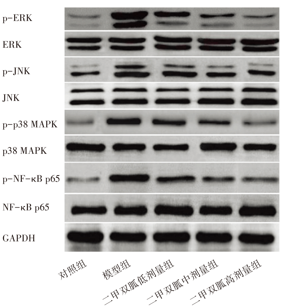
| 组别 | n | p-ERK | p-JNK | p-p38 MAPK | p-NF-κB p65 |
|---|---|---|---|---|---|
| 对照组 | 10 | 0.21±0.03 | 0.19±0.02 | 0.26±0.04 | 0.28±0.03 |
| 模型组 | 10 | 1.07±0.09a | 0.94±0.08a | 0.84±0.07a | 0.91±0.09a |
| 二甲双胍低剂量组 | 10 | 0.82±0.06ab | 0.76±0.07ab | 0.71±0.06ab | 0.77±0.07ab |
| 二甲双胍中剂量组 | 10 | 0.69±0.05abc | 0.57±0.05abc | 0.57±0.05abc | 0.58±0.06abc |
| 二甲双胍高剂量组 | 10 | 0.43±0.04abcd | 0.38±0.04abcd | 0.42±0.04abcd | 0.46±0.05abcd |
| F | 324.005 | 253.897 | 168.355 | 149.563 | |
| P | <0.001 | <0.001 | <0.001 | <0.001 |
| [1] | Siddiqui S, Mateen S, Ahmad R, et al. A brief insight into the etiology, genetics, and immunology of polycystic ovarian syndrome (PCOS)[J]. J Assist Reprod Genet, 2022, 39(11):2439-2473. doi: 10.1007/s10815-022-02625-7. |
| [2] | Armanini D, Boscaro M, Bordin L, et al. Controversies in the Pathogenesis, Diagnosis and Treatment of PCOS: Focus on Insulin Resistance, Inflammation, and Hyperandrogenism[J]. Int J Mol Sci, 2022, 23(8):4110. doi: 10.3390/ijms23084110. |
| [3] |
Liu K, He X, Huang J, et al. Short-chain fatty acid-butyric acid ameliorates granulosa cells inflammation through regulating METTL3-mediated N6-methyladenosine modification of FOSL2 in polycystic ovarian syndrome[J]. Clin Epigenetics, 2023, 15(1):86. doi: 10.1186/s13148-023-01487-9.
pmid: 37179374 |
| [4] |
Guo Y, Zhang H, Lv Z, et al. Up-regulated CD38 by daphnetin alleviates lipopolysaccharide-induced lung injury via inhibiting MAPK/NF-κB/NLRP3 pathway[J]. Cell Commun Signal, 2023, 21(1):66. doi: 10.1186/s12964-023-01041-3.
pmid: 36998049 |
| [5] |
Zhao X, Li M, Lu Y, et al. Sirt1 inhibits macrophage polarization and inflammation in gouty arthritis by inhibiting the MAPK/NF-κB/AP-1 pathway and activating the Nrf2/HO-1 pathway[J]. Inflamm Res, 2024, 73(7):1173-1184. doi: 10.1007/s00011-024-01890-9.
pmid: 38739197 |
| [6] |
Zhao H, Xing C, Zhang J, et al. Comparative efficacy of oral insulin sensitizers metformin, thiazolidinediones, inositol, and berberine in improving endocrine and metabolic profiles in women with PCOS: a network meta-analysis[J]. Reprod Health, 2021, 18(1):171. doi: 10.1186/s12978-021-01207-7.
pmid: 34407851 |
| [7] | Kukaev E, Kirillova E, Tokareva A, et al. Impact of Gut Microbiota and SCFAs in the Pathogenesis of PCOS and the Effect of Metformin Therapy[J]. Int J Mol Sci, 2024, 25(19):10636. doi: 10.3390/ijms251910636. |
| [8] | Balasubramanian A, Pachiappan S, Mohan S, et al. Therapeutic exploration of polyherbal formulation against letrozole induced PCOS rats: A mechanistic approach[J]. Heliyon, 2023, 9(5):e15488. doi: 10.1016/j.heliyon.2023.e15488. |
| [9] | Dabravolski SA, Nikiforov NG, Eid AH, et al. Mitochondrial Dysfunction and Chronic Inflammation in Polycystic Ovary Syndrome[J]. Int J Mol Sci, 2021, 22(8):3923. doi: 10.3390/ijms22083923. |
| [10] | 袁山政, 张燕雨, 翟亚悦, 等. 循环炎症因子与PCOS风险之间的关联评估:一项双样本孟德尔随机化研究[J]. 生殖医学杂志, 2024, 33(9):1214-1220. doi: 10.3969/j.issn.1004-3845.2024.09.013. |
| [11] | 刘聪, 杜宇坤, 折慧芝, 等. 二甲双胍联合枸橼酸氯米芬治疗对PCOS合并不孕症患者胰岛素、HOMA-IR水平及卵巢排卵功能的影响[J]. 生殖医学杂志, 2025, 34(4):430-435. doi: 10.3969/j.issn.1004-3845.2025.04.002. |
| [12] | Kumari R, Singh A, Banerjee BD, et al. Impact of Metformin Therapy on Chronic Inflammatory Markers Serum Fractalkine and CRP Levels in Polycystic Ovary Syndrome: A Pilot Study on Chronic Inflammatory Markers and Clinical Outcomes[J]. J Obstet Gynaecol India, 2025, 75(2):115-121. doi: 10.1007/s13224-025-02100-0. |
| [13] |
Yousuf SD, Ganie MA, Urwat U, et al. Oral contraceptive pill (OCP) treatment alters the gene expression of intercellular adhesion molecule-1 (ICAM-1), tumor necrosis factor-α (TNF-α), monocyte chemoattractant protein-1 (MCP-1) and plasminogen activator inhibitor-1 (PAI-1) in polycystic ovary syndrome (PCOS) women compared to drug-naive PCOS women[J]. BMC Womens Health, 2023, 23(1):68. doi: 10.1186/s12905-023-02187-5.
pmid: 36793022 |
| [14] | Bansal B, Thazhuthadath Kishore A, Kathiresan S, et al. A Systematic Review of Inflammatory Markers in Polycystic Ovary Syndrome (PCOS) and Meta-Analysis of Interleukin-6 (IL-6) in Case-Control Studies[J]. Cureus, 2025, 17(4):e82350. doi: 10.7759/cureus.82350. |
| [15] | 朱松楠, 张红媛, 李艳. 血清NLRP3、IL-18水平与多囊卵巢综合征患者不孕的关系[J]. 中国现代医学杂志, 2024, 34(13):1-6. doi: 10.3969/j.issn.1005-8982.2024.13.001. |
| [16] | 符山花, 包利利, 赵达, 等. 抑制NLRP3炎症小体激活可调节自噬改善多囊卵巢综合征颗粒细胞凋亡[J]. 中国免疫学杂志, 2024, 40(8):1646-1652. doi: 10.3969/j.issn.1000-484X.2024.08.013. |
| [17] |
Liu Z, Wang RH, Wang KH. Formononetin ameliorates polycystic ovary syndrome through suppressing NLRP3 inflammasome[J]. Mol Med, 2025, 31(1):27. doi: 10.1186/s10020-025-01092-x.
pmid: 39871124 |
| [18] | 李春颖, 尹燕燕, 季梦垚, 等. 基于MAPK/NF-κB信号通路探讨调气止咳方对咳嗽变异性哮喘大鼠气道炎症的影响及机制[J]. 中药新药与临床药理, 2024, 35(7):1008-1015. doi: 10.19378/j.issn.1003-9783.2024.07.009. |
| [19] | 柯玲玲, 王敏, 高洁, 等. STAT1对肺结核大鼠巨噬细胞凋亡及MAPK/NF-κB信号通路的机制研究[J]. 解剖学研究, 2024, 46(2):115-122. doi: 10.20021/j.cnki.1671-0770.2024.02.04. |
| [20] | 曹静, 刘海波, 安琪, 等. 二甲双胍缓解小鼠放射性皮炎引起的病理性疼痛:基于抑制p38 MAPK/NF-κB信号通路[J]. 南方医科大学学报, 2023, 43(10):1815-1820. doi: 10.12122/j.issn.1673-4254.2023.10.22. |
| [21] | 黄辉, 于大海, 黄炫赓, 等. 二甲双胍对糖基化终末产物诱导牙周膜成纤维细胞凋亡及p38 MAPK/NF-κB通路的影响[J]. 山东医药, 2021, 61(14):19-23. doi: 10.3969/j.issn.1002-266X.2021.14.005. |
| [1] | HAN Zhao-xun, ZHOU Ye. Research Progress on Oxidative Stress Mechanisms and Antioxidant Therapy in Endometriosis [J]. Journal of International Obstetrics and Gynecology, 2026, 53(2): 125-130. |
| [2] | CAI Hao-qin, WANG Yu, ZHANG Xi, YU Jian-nan, SHI Bai-chao, WU Xiao-ke. Potential Therapeutic Effects of Dendrobium Officinale Polysaccharide in Polycystic Ovary Syndrome [J]. Journal of International Obstetrics and Gynecology, 2026, 53(2): 131-136. |
| [3] | ZHAO Jia-run, ZHENG Shu-chang, WANG Yi-di, WANG Cheng-xia, HE Jun-qin. Immune-Inflammatory Thrombotic Mechanism of Recurrent Spontaneous Abortion Caused by Antiphospholipid Syndrome [J]. Journal of International Obstetrics and Gynecology, 2026, 53(1): 103-107. |
| [4] | DING Ning, HAN Yan-hua, WANG Hao-tian, SUN Chang, KUANG Hong-ying. The Mechanism of Exosomal MicroRNAs in Polycystic Ovary Syndrome [J]. Journal of International Obstetrics and Gynecology, 2026, 53(1): 73-77. |
| [5] | Xiemuxinuer • Simayi, HUANG Ya-nan, ZHANG Man-li, HAN Rui. Advances in Adipose-Tissue Immunity and Metabolism in Polycystic Ovary Syndrome [J]. Journal of International Obstetrics and Gynecology, 2026, 53(1): 78-84. |
| [6] | LIU Yin, SONG Dian-rong. Research Progress on the Treatment of Polycystic Ovary Syndrome Based on Improving Insulin Resistance [J]. Journal of International Obstetrics and Gynecology, 2025, 52(6): 601-605. |
| [7] | ZHAO Rui-kun, ZHOU Qin. Research Progress on the Pathogenesis of Endometrial Polyps [J]. Journal of International Obstetrics and Gynecology, 2025, 52(6): 624-628. |
| [8] | LENG Ya-wen, WANG Yu, WU Xiao-ke. Research Progress on the Treatment of Polycystic Ovary Syndrome with Human Umbilical Cord Mesenchymal Stem Cell [J]. Journal of International Obstetrics and Gynecology, 2025, 52(5): 527-533. |
| [9] | ZENG Hui-fang, DOU Zhen, LI Yu-xin, JIANG Xin-yu, XIA Tian. Research Progress on Glucose and Lipid Metabolism in Decidualization of Endometrial Stromal Cells in Patients with PCOS [J]. Journal of International Obstetrics and Gynecology, 2025, 52(5): 534-539. |
| [10] | CHEN Yi-bing, TU Jing-yan, TANG Yi-ming, LIN Xiao-yang, LIU Yan-duo, HAN Qing. Research Progress on Serum Iron and Ferroptosis in Preeclampsia [J]. Journal of International Obstetrics and Gynecology, 2025, 52(4): 361-365. |
| [11] | SHAO Meng-yu, MA Sai-hua, GONG Zheng, ZHAO Xiao-li, ZHAO Zhi-mei. Pathogenesis of Endometriosis Complicated with Chronic Endometritis and Its Impact on Reproduction [J]. Journal of International Obstetrics and Gynecology, 2025, 52(3): 241-245. |
| [12] | XU Shu-ying, XU Hai-peng, WANG Li-na, ZHANG Yang. Relationship between Zinc and Polycystic Ovary Syndrome [J]. Journal of International Obstetrics and Gynecology, 2025, 52(2): 217-221. |
| [13] | YAN Hui-bo, ZHANG Lin. Analysis of the Disease Burden and Projections of Polycystic Ovary Syndrome in China and Globally from 1990 to 2021 [J]. Journal of International Obstetrics and Gynecology, 2025, 52(2): 228-233. |
| [14] | CHEN Shu-lin, QIAO Qiao. Relationship between Vaginal Epithelial Injury Repair and Microecological Environment [J]. Journal of International Obstetrics and Gynecology, 2025, 52(1): 52-56. |
| [15] | ZHANG Dong, WANG Zheng, LI Kai, BIAN Wen-li, GAO Zhi-hua. A Case of Severe Spontaneous Ovarian Hyperstimulation Syndrome in Non-Pregnancy [J]. Journal of International Obstetrics and Gynecology, 2025, 52(1): 79-83. |
| Viewed | ||||||
|
Full text |
|
|||||
|
Abstract |
|
|||||