国际妇产科学杂志 ›› 2026, Vol. 53 ›› Issue (1): 85-88.doi: 10.12280/gjfckx.20251335

• 普通妇科疾病及相关研究: 病例报告 • 上一篇    下一篇

子宫腺肌病合并肌壁间脓肿一例

吕寒雪(), 徐金颖, 刘华()   

  1. 261000 山东省潍坊市,山东第二医科大学附属医院(临床医学院)(吕寒雪,徐金颖);山东第二医科大学附属医院妇科(刘华)
  • 收稿日期:2025-11-28 出版日期:2026-02-15 发布日期:2026-03-11
  • 通讯作者: 刘华 E-mail:345208565@qq.com

Adenomyosis Complicated with Intramural Uterine Abscess: A Case Report

LYU Han-xue(), XU Jin-ying, LIU Hua()   

  1. Affiliated Hospital of Shandong Second Medical University (School of Clinical Medicine), Weifang 261000, Shandong Province, China (LYU Han-xue, XU Jin-ying); Department of Gynecology, Affiliated Hospital of Shandong Second Medical University, Weifang 261000, Shandong Province, China (LIU Hua)
  • Received:2025-11-28 Published:2026-02-15 Online:2026-03-11
  • Contact: LIU Hua E-mail:345208565@qq.com

摘要:

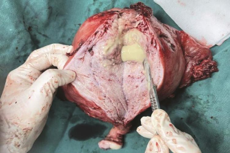
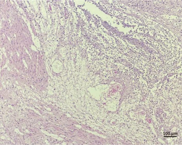
报告1例50岁子宫腺肌病合并子宫肌壁间脓肿及输卵管积脓患者。患者因腹痛、发热入院,检查提示重症感染(白细胞47.05×10⁹/L)及子宫显著增大。初始经验性抗生素(亚胺培南西司他丁钠)治疗后感染指标部分下降,但腹痛加剧并出现腹腔积液,怀疑脓肿破裂。急行经腹全子宫切除术+双侧附件切除术+盆腔粘连松解术+肠粘连分解术,术中见盆腔脓性分泌物,术后剖视子宫标本证实肌壁间存在多发脓腔,病理诊断符合子宫腺肌瘤伴感染及脓肿形成。术后根据病原学结果继续抗感染及支持治疗,患者感染得到控制。本例提示子宫腺肌病可能成为盆腔感染扩散、形成深部肌壁间脓肿的基础。对于此类复杂性感染,在强效抗生素治疗下若病情反复或疑有脓肿破裂,采取手术治疗控制感染源是成功救治的关键。

关键词: 子宫腺肌病, 脓肿, 宫腔积脓, 盆腔炎性疾病, 治疗

Abstract:

We report a case of a 50-year-old female patient with adenomyosis complicated by an intramural uterine abscess and pyosalpinx. The patient was admitted due to abdominal pain and fever. Laboratory examinations indicated severe infection (leukocyte count: 47.05×10⁹/L) and imaging revealed significant uterine enlargement. Following initial empirical antibiotic therapy (imipenem/cilastatin sodium), infectious parameters partially decreased. However, the abdominal pain intensified and ascites developed, raising suspicion of abscess rupture. Emergency exploratory laparotomy with total abdominal hysterectomy, bilateral salpingo-oophorectomy, pelvic adhesiolysis and enterolysis was performed. Intraoperative findings included purulent exudate in the pelvic cavity. Postoperative dissection of the uterine specimen confirmed multiple abscess cavities within the myometrium. Histopathological diagnosis was consistent with adenomyoma with superimposed infection and abscess formation. Postoperatively, based on etiological results, targeted antibiotic therapy and supportive care were continued, leading to successful infection control. This case highlights that adenomyosis may serve as a nidus for pelvic infection dissemination and the formation of deep intramural abscesses. For such complicated infections, if clinical condition deteriorates or abscess rupture is suspected despite potent antibiotic therapy, surgical intervention to control the source of infection is critical for successful management.

Key words: Adenomyosis, Abscess, Pyometra, Pelvic inflammatory disease, Therapy