Journal of International Obstetrics and Gynecology ›› 2021, Vol. 48 ›› Issue (4): 434-437.doi: 10.12280/gjfckx.20200825
• Gynecological Disease & Related Research Original Article • Previous Articles Next Articles
JIA Hong-jing, DENG Xue-dong, CHEN Xiao-min, CAO Jiao-jiao, MA Lei, LU Bing( )
)
Received:2020-09-07
Published:2021-08-15
Online:2021-09-01
Contact:
LU Bing
E-mail:lulum18@163.com
JIA Hong-jing, DENG Xue-dong, CHEN Xiao-min, CAO Jiao-jiao, MA Lei, LU Bing. Application of Three- and Four-Dimensional Transperineal Ultrasound in Diagnosis of Pelvic Organ Prolapse by Measuring Area of Levator Hiatus[J]. Journal of International Obstetrics and Gynecology, 2021, 48(4): 434-437.
Add to citation manager EndNote|Ris|BibTeX
| 组别 | n | 静息状态 | Valsalva-3D | Valsalva-4D | t′ | P′ |
|---|---|---|---|---|---|---|
| POP组 | 84 | 13.97±2.63 | 23.79±4.81 | 23.61±4.79 | 1.020 | 0.311 |
| 对照组 | 69 | 11.62±1.97 | 15.59±2.48 | 15.70±2.53 | 0.888 | 0.378 |
| t | 6.303 | 13.584 | 13.064 | |||
| P | 0.000 | 0.000 | 0.000 |
| 组别 | n | 静息状态 | Valsalva-3D | Valsalva-4D | t′ | P′ |
|---|---|---|---|---|---|---|
| POP组 | 84 | 13.97±2.63 | 23.79±4.81 | 23.61±4.79 | 1.020 | 0.311 |
| 对照组 | 69 | 11.62±1.97 | 15.59±2.48 | 15.70±2.53 | 0.888 | 0.378 |
| t | 6.303 | 13.584 | 13.064 | |||
| P | 0.000 | 0.000 | 0.000 |
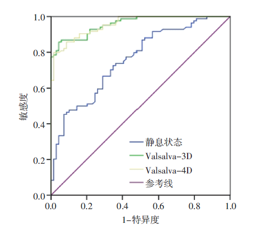
| 组别 | 截断值 (cm2) | AUC | 准确度 (%) | 敏感度 (%) | 特异度 (%) |
|---|---|---|---|---|---|
| 静息状态 | 14.29 | 0.759 | 66.67 | 45.24 | 92.75 |
| Valsalva-3D | 19.29 | 0.959a | 90.20 | 85.71 | 95.65 |
| Valsalva-4D | 19.90 | 0.956ab | 88.24 | 79.76 | 98.55 |
| 组别 | 截断值 (cm2) | AUC | 准确度 (%) | 敏感度 (%) | 特异度 (%) |
|---|---|---|---|---|---|
| 静息状态 | 14.29 | 0.759 | 66.67 | 45.24 | 92.75 |
| Valsalva-3D | 19.29 | 0.959a | 90.20 | 85.71 | 95.65 |
| Valsalva-4D | 19.90 | 0.956ab | 88.24 | 79.76 | 98.55 |
| [1] | 朱兰, 郎景和. 女性盆底学[M]. 北京: 人民卫生出版社, 2014: 113. |
| [2] |
毛卉, 黄程胜. 生物力学在女性盆底功能障碍性疾病中的应用[J]. 国际妇产科学杂志, 2019, 46(2):212-215. doi: 10.3969/j.issn.1674-1870.2019.02.025.
doi: 10.3969/j.issn.1674-1870.2019.02.025 |
| [3] |
李志毅, 朱兰, 徐涛, 等. 中国城市地区女性盆腔器官脱垂临床流行病学调查[J]. 中华医学杂志, 2019, 99(11):857-861. doi: 10.3760/cma.j.issn.0376-2491.2019.11.012.
doi: 10.3760/cma.j.issn.0376-2491.2019.11.012 |
| [4] | 张新玲. 实用盆底超声诊断学[M]. 北京: 人民卫生出版社, 2018: 1. |
| [5] |
Abdool Z, Dietz HP, Lindeque BG. Ethnic differences in the levator hiatus and pelvic organ descent: a prospective observational study[J]. Ultrasound Obstet Gynecol, 2017, 50(2):242-246. doi: 10.1002/uog.17297.
doi: 10.1002/uog.17297 pmid: 27607844 |
| [6] |
吴曼丽, 林欣, 王旭东, 等. 肛提肌裂孔与盆腔器官脱垂量化分期及脱垂症状的相关性分析[J]. 中华超声影像学杂志, 2020, 29(8):700-705. doi: 10.3760/cma.j.cn131148-20200325-00229.
doi: 10.3760/cma.j.cn131148-20200325-00229 |
| [7] |
吴晓翔, 张凤玲, 刘秀平, 等. 经会阴部超声检查在绝经后女性肛提肌裂孔形变与盆底功能障碍性疾病的临床价值[J]. 重庆医学, 2018, 47(18):2504-2506. doi: 10.3969/j.issn.1671-8348.2018.18.030.
doi: 10.3969/j.issn.1671-8348.2018.18.030 |
| [8] |
王晶晶, 张枢书, 黄晓玲. 超声评价肛提肌及其裂孔在女性盆腔器官脱垂中的进展[J]. 国际妇产科学杂志, 2019, 46(3):351-354. doi: 10.3969/j.issn.1674-1870.2019.03.026.
doi: 10.3969/j.issn.1674-1870.2019.03.026 |
| [9] |
Handa VL, Roem J, Blomquist JL, et al. Pelvic organ prolapse as a function of levator ani avulsion, hiatus size, and strength[J]. Am J Obstet Gynecol, 2019, 221(1):41.e1-41.e7. doi: 10.1016/j.ajog.2019.03.004.
doi: 10.1016/j.ajog.2019.03.004 |
| [10] |
Margulies RU, Hsu Y, Kearney R, et al. Appearance of the levator ani muscle subdivisions in magnetic resonance images[J]. Obstet Gynecol, 2006, 107(5):1064-1069. doi: 10.1097/01.AOG.0000214952.28605.e8.
doi: 10.1097/01.AOG.0000214952.28605.e8 pmid: 16648412 |
| [11] |
Zhu YC, Deng SH, Jiang Q, et al. Correlation Between Delivery Mode and Pelvic Organ Prolapse Evaluated by Four-Dimensional Pelvic Floor Ultrasonography[J]. Med Sci Monit, 2018, 24:7891-7897. doi: 10.12659/MSM.911343.
doi: 10.12659/MSM.911343 |
| [12] |
徐英姿, 唐海林, 冯泽阳. 盆底超声检查观察二次自然分娩对女性盆底结构的近期影响[J]. 中华医学超声杂志(电子版), 2018, 15(3):218-222. doi: 10.3877/cma.j.issn.1672-6448.2018.03.011.
doi: 10.3877/cma.j.issn.1672-6448.2018.03.011 |
| [13] |
曹晓燕, 齐艳, 赵华云. 经会阴四维超声成像检测产后盆底功能障碍性疾病的应用价值[J]. 临床超声医学杂志, 2018, 20(7):468-471. doi: 10.3969/j.issn.1008-6978.2018.07.013.
doi: 10.3969/j.issn.1008-6978.2018.07.013 |
| [14] |
Siafarikas F, Staer-Jensen J, Braekken IH, et al. Learning process for performing and analyzing 3D/4D transperineal ultrasound imaging and interobserver reliability study[J]. Ultrasound Obstet Gynecol, 2013, 41(3):312-317. doi: 10.1002/uog.11192.
doi: 10.1002/uog.11192 pmid: 22605574 |
| [15] |
叶婷婷, 王慧芳, 陈华, 等. 盆底超声智能识别及半自动测量泌尿生殖裂孔的实用性研究[J]. 中华超声影像学杂志, 2019, 28(3):256-260. doi: 10.3760/cma.j.issn.1004-4477.2019.03.013.
doi: 10.3760/cma.j.issn.1004-4477.2019.03.013 |
| [16] |
鲁蓉, 张瑜, 俞亚平. 超声测量肛提肌裂孔面积在子宫脱垂诊断中的应用[J]. 中华医学杂志, 2019, 99(29):2315-2318. doi: 10.3760/cma.j.issn.0376-2491.2019.29.014.
doi: 10.3760/cma.j.issn.0376-2491.2019.29.014 |
| [17] |
肖汀, 张新玲, 杨丽新, 等. 超声测量肛提肌裂孔面积在女性压力性尿失禁诊断中的应用[J]. 中国医学影像技术, 2016, 32(9):1419-1422. doi: 10.13929/j.1003-3289.2016.09.026.
doi: 10.13929/j.1003-3289.2016.09.026 |
| [18] |
Dietz HP. Ultrasound in the assessment of pelvic organ prolapse[J]. Best Pract Res Clin Obstet Gynaecol, 2019, 54:12-30. doi: 10.1016/j.bpobgyn.2018.06.006.
doi: 10.1016/j.bpobgyn.2018.06.006 |
| [1] | ZHANG Yong-qing, CHEN Zheng-yun, CHEN Lu-ping, YAN Guo-hui, CHEN Dan-qing. Two Cases of Term Angular Pregnancy Identified during Cesarean Section [J]. Journal of International Obstetrics and Gynecology, 2025, 52(2): 153-157. |
| [2] | LIN Huan-yu, YU Min, LU Xu-hong. Research Progress on High-Risk Factors for Postpartum Pelvic Floor Dysfunction [J]. Journal of International Obstetrics and Gynecology, 2024, 51(6): 620-623. |
| [3] | SHAO Hui, WANG Lu, CHEN Lu-jia, DONG Rui-jia, TANG Jie-ying, LIAO Xue-yin, YANG Jian-min, LI Wei-wei. Prospective Randomized Controlled Clinical Study on the Efficacy of A Novel Temperature-Controlled Radiofrequency Treatment for Postpartum Vaginal Laxity [J]. Journal of International Obstetrics and Gynecology, 2024, 51(5): 509-514. |
| [4] | TAN Mi, TAN Qing-qing. Non-Surgical Treatment Methods for Pelvic Floor Dysfunction [J]. Journal of International Obstetrics and Gynecology, 2024, 51(4): 401-405. |
| [5] | ZHANG Yan, HU Meng-ying, WANG Hua, DONG Qu-long. Progress in The Application of Ultrasound-Guided High Intensity Focused Ultrasound in Gynecological and Obstetric Diseases [J]. Journal of International Obstetrics and Gynecology, 2024, 51(4): 406-411. |
| [6] | WANG Qiu-ming, WANG Rui-li, WU Hai-ying, WANG Li. Intrafetal Radiofrequency Ablation for Fetal Pulmonary Sequestration with Edema and Pleural Effusion: A Case Report [J]. Journal of International Obstetrics and Gynecology, 2024, 51(4): 477-480. |
| [7] | ZHANG Chun-shuang, DONG Xiao-zhen, ZHOU Chang-rong, WANG Yi-shan, LI He-zhou. Intrauterine Treatment of Fetal Hydrothorax Combined with Hydrops Complicating Mirror Syndrome:A Case Report [J]. Journal of International Obstetrics and Gynecology, 2024, 51(3): 357-360. |
| [8] | LIU Shu-jie, ZHANG Hai-Yan. Research Progress of Laparoscopic Lateral Suspension with Mesh and Its Modified Operation in the Treatment of Pelvic Organ Prolapse [J]. Journal of International Obstetrics and Gynecology, 2024, 51(2): 121-127. |
| [9] | WANG Qi-qin, WANG Xiang-lian, PAN Si-yi, WANG Xiu-li. Research Progress of Premenopausal Endometrial Polyps, Postmenopausal Endometrial Polyps and Tamoxifen-Associated Endometrial Polyps [J]. Journal of International Obstetrics and Gynecology, 2024, 51(2): 137-141. |
| [10] | WANG Xiao-li, CHEN Yi, ZHENG Xin-chun, CAO Yan-hua. A Case Report of Atypical Accessory Cavitated Uterine Malformation [J]. Journal of International Obstetrics and Gynecology, 2024, 51(2): 148-151. |
| [11] | YANG Qiong, XU Xiao-yan. Analysis of Maternal and Infant Outcomes of Septate Uterus in Late Pregnancy by Cesarean Section [J]. Journal of International Obstetrics and Gynecology, 2024, 51(2): 181-183. |
| [12] | LUO Ting, LIU Bo, ZHOU Zhong-min, HOU Shu-hui, LIU Jin-yu, PENG Mei. A Case Report of Triploid Stillbirth with Cyclopia [J]. Journal of International Obstetrics and Gynecology, 2024, 51(2): 203-205. |
| [13] | XUE Feng-qin, ZHAO Shu-rui, ZHAO Ye. Advances in the Anatomical and Histological Characteristics of the Human Uterosacral Ligament and Related Biomechanical Studies [J]. Journal of International Obstetrics and Gynecology, 2023, 50(6): 606-612. |
| [14] | ZHU Jing, XIA Zhi-jun. Human Vaginal Fibroblast Dysfunction in Pelvic Organ Prolapse [J]. Journal of International Obstetrics and Gynecology, 2023, 50(6): 613-617. |
| [15] | MA Shuai, XU Ying, WANG Min, LI Jie. Advances in the Study of Fetal Intracranial Hemorrhage [J]. Journal of International Obstetrics and Gynecology, 2023, 50(5): 481-485. |
| Viewed | ||||||
|
Full text |
|
|||||
|
Abstract |
|
|||||